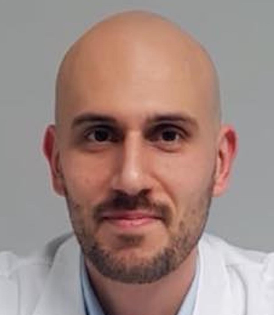
Dr. Riccardo Pampena


M.D. Freelance Dermatologist,
Centro Oncologico ad Alta Tecnologia Diagnostica, Azienda Unità Sanitaria Locale, IRCCS di Reggio Emilia, Italy
Ex vivo fluorescence confocal microscopy for Mohs surgery of basal cell carcinomas
AUTHOR: Riccardo Pampena MD
Frozen histological sections are currently used for intra-operative margin assessment during Mohs surgery. Fluorescence confocal microscopy (FCM) is a new tool, which offers a promising and faster alternative to frozen histology.
We prospectively evaluated in a clinical setting, the accuracy of FCM as compared to frozen sections in BCC’s margin assessment.
Patients with BCC, scheduled for Mohs surgery were prospectively enrolled. Freshly-excised surgical specimens were first examined through FCM and then frozen sections were evaluated. Permanent sections were finally obtained, in order to validate our sample technique. A blind re-evaluation was also performed for discordant cases between FCM and frozen sections. Sensitivity and specificity levels, as well as positive and negative predicting values were calculated and ROC curves were generated.
A total of 127 BCCs in as many patients (40.2% females) were enrolled. A total number of 753 sections were examined. All BCCs were located on the head and neck area. When evaluating the performance of FCM as compared to frozen sections 79.8% sensitivity, 95.8% specificity, 80.5% positive predicting and 95.7% negative predicting values were found (Area Under the Curve: .88, 95%CI .84-.92; P<.001). A total of 49 discordant cases between FCM and frozen sections evaluations were blindly re-evaluated, of which 24 were false positive and 25 false negative. The performance of FCM and frozen sections was also evaluated according to the final histopathological assessment.
In conclusion we found high levels of accuracy for FCM as compared to frozen sections evaluation, in intra-operative BCC’s margin assessment during Mohs surgery. Some technical issues still prevent a wide use of this technique, but new upcoming devices promise to overcome these limitations.
SHORT CV
Dr. Riccardo Pampena is a board-certified Dermatologist specialized in the diagnosis and treatment of skin cancers.
He obtained his degree in Surgery and Medicine (MD) from the Catholic University of the Sacred Heart of Rome in 2011 with full mark; then, he became a board-certified Dermatologist (full mark) in 2016 at the Department of Dermatology and Venereology of the Sapienza University of Rome.
Since 2011 he practices research mainly in the field of psoriasis and non-invasive diagnosis in dermato-oncology, paying particular attention to the use of non-invasive diagnostic methods, such as dermoscopy and confocal microscopy for the in vivo and ex vivo study. Since November 2016, Dr. Pampena has been working at the Skin Cancer Unit of the Arcispedale Santa Maria Nuova of Reggio Emilia, a third-level referral center for skin tumors diagnosis and management and recently became a Board Member of the International Dermoscopy Society.
Furthermore, he’s actively involved, as a statistician, in scientific studies and was the first author of a meta-analysis on “Nevus associated melanoma” published on the prestigious Journal of the American Academy of Dermatology in 2017.
Contactează operatorul PDI 2019
Operatorul PDI 2019
Adresa: Str. A. Panu nr. 13, Iasi
Tel.: 0332.40.88.00-05
E-mail: inscrieri@pdi.ro/2019
Website: www.eventernet.ro
