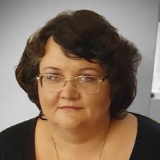
Prof. Univ. Dr. Zurac Sabina


Prof. Dr. Zurac Sabina
Universitatea de Medicină şi Farmacie “Carol Davila” Bucureşti
Spitalului Clinic Colentina, București
Inteligența artificială în histopatologie: Experiența Spitalului Clinic Colentina și Zaya Artificial Intelligence
Autori: Sabina Zurac1,2,3*, Cristiana Popp1,3*, M Trascau3,4*, C Mogodici3*, Luciana Nichita1,2,3, Mirela Cioplea1,2,3, T Poncu3, B Ceachi3, Liana Sticlaru1,2,3, Alexandra Cioroianu1,2,3, M Busca1,3, Oana Stefan1, Irina Tudor1, A Voicu3, Daliana Stanescu3, Alexandra Bastian1,2
*autori cu contributie egala
1 Spitalul Clinic Colentina
2 Universitatea de Medicină și Farmacie Carol Davila Bucuresti
3 Zaya Artificial Intelligence SRL
4 Universitatea Politehnică Bucuresti
Inteligența artificială, tuberculoza, mycobacterium tuberculosis, Ziehl Nielsen
Tuberculoza reprezintă prima cauză de deces prin boală infecțioasă. Ideal, diagnosticul TBC se bazează pe identificarea Mycobacteriilor. Deoarece Mycobacterium tuberculosis este o bacterie de 2-4/0,2-0,5 microni, căutarea ei în lamele colorate Ziehl Nielsen (ZN) cu obiectiv microscopic 40X (diametru 0,5 mm) durează ore; inteligența artificială poate oferi soluții viabile în acest domeniu.
Echipa noastră include patologi, ingineri de machine learning și experți în IT și economie. Setul de date de antrenare include 500.000 de casete de delimitare pozitive și 1.000.000 negative provenite din lamele ZN a 81 de blocuri de parafină (21 pozitive, 60 negative); toate lamele au fost scanate folosind scanere manuale și automate; 7 patologi au notat zone pozitive și negative. Echipa de machine learning a propus mai multe tehnici de augmentare a imaginii cuplate cu diferite arhitecturi de computer vision. Setul de date de testare include 23 de lame ZN pozitive și 23 negative (46 de blocuri); lamele au fost scanate integral (whole slide images – WSI). 4 echipe de patologi au evaluat separat WSI-urile și lamele; WSI-urile au fost încărcate pentru analiză automată.
Analiza automată a WSI generează un raport care indică zonele susceptibile de a prezenta micobacterii. Arhitectura noastră prezinta rezultate foarte bune (0.9020 pe curba ROC). Rezultatele testului final (mașină versus om care evaluează WSI, mașină versus om care evaluează lame și om care evaluează WSI versus lame) arată acuratețe peste 90% în toate cazurile.
Software-ul bazat pe inteligență artificială pentru analiza automată a lamelor colorate ZN prezintă rezultate fiabile, oferind o economie extraordinară în timpul și efortul patologului.
Artificial Intelligence in histopathology: Colentina University Hospital and Zaya Artificial Intelligence experience
Autori: Sabina Zurac1,2,3*, Cristiana Popp1,3*, M Trascau3,4*, C Mogodici3*, Luciana Nichita1,2,3, Mirela Cioplea1,2,3, T Poncu3, B Ceachi3, Liana Sticlaru1,2,3, Alexandra Cioroianu1,2,3, M Busca1,3, Oana Stefan1, Irina Tudor1, A Voicu3, Daliana Stanescu3, Alexandra Bastian1,2
*Authors with equal contribution
1 Colentina University Hospital
2 University of Medicine and Pharmacy Carol Davila Bucharest
3 Zaya Artificial Intelligence SRL
4 University of Polytechnics Bucharest
Artificial intelligence, tuberculosis, mycobacterium tuberculosis, Ziehl Nielsen
Tuberculosis represents the first cause of death by infectious diseases. Ideally, TB diagnosis relies on Mycobacteria identification. Since Mycobacterium tuberculosis is a 2-4/0.2-0.5 microns bacterium, its searched in Ziehl Nielsen (ZN) stained-slides with 40X microscopic objective (0.5mm diameter) takes hours; artificial intelligence may offer viable solutions in this field.
Our team includes pathologists from Colentina University Hospital, machine learning engineers from University of Polytechnics Bucharest and experts in IT and economics from Zaya Artificial Intelligence SRL. Our training data set includes 500.000 positive and 1.000.000 negative bounding boxes originating in ZN-stained slides of 81 paraffin blocks (21 positive, 60 negative); all the slides were scanned using manual and automated slide scanners; 7 pathologists annotated positive and negative areas. The machine learning team has proposed several image augmentation techniques coupled with different custom computer vision architectures. Our testing data set includes 23 positive and 23 negative ZN-stained slides (from 46 paraffin blocks); the slides were scanned as whole slide images (WSIs). 4 teams of pathologists separately evaluated the WSIs and the original slides; the WSIs were sent for automatic analysis.
WSIs automatic analysis was followed by a report indicating areas more likely to present Mycobacteria. Our architecture showed very good results (0.9020 on ROC curve). The results of final test (machine versus man evaluating WSIs, machine versus man evaluating slides and man evaluating WSIs versus slides) showed accuracy over 90% in all cases.
AI-based software for automated analysis of ZN-stained slides shows reliable results, offering a tremendous economy in pathologist’s time&effort.
Scurt CV
Professor of Pathology in Faculty of Dental Medicine, University of Medicine and Pharmacy “Carol Davila” Bucharest, senior pathologist and head of the Department of Pathologicy Colentina University Hospital, Bucharest, Romania.
Specializations in the field of dermatopathology (Austria 2012), molecular biology (Italy 2012), digestive pathology (Bucharest 2016, 2015, 2011, 2010, 2009 and 2008), oral pathology (the Netherlands 2005-2006), immunohistochemistry (Bucharest 2004; Greece 2001 ), lymphoid pathology (Greece 2001), pulmonary pathology (Italy 1999), soft tissue pathology (Greece 1997), liver pathology (Greece 1996)
Member of the European Society of Pathology, International Academy of Pathology, International Society of Dermatopathology, Romanian Society of Dermatooncology, Romanian Association of Immunodermatology, European Academy of Dermatovenerology
Author of over 330 scientific papers presented at national and international congresses and symposiums, over 125 articles published in specialized magazines, 23 specialized chapters / monographs. H-index (WOS all data bases) 10; citations: 460.
Researcher / manager / project manager in 16 research projects
Areas of interest: dermatopathology, oral pathology, infectious pathology, soft tissue tumors
Contactează operatorul PDI 2022
Operatorul PDI 2022
![]()
Adresa: Str. A. Panu nr. 13, Iasi
Tel.: 0332.40.88.00-05
E-mail: contact@pdi.ro
Website: www.eventernet.ro
Parteneri Media
